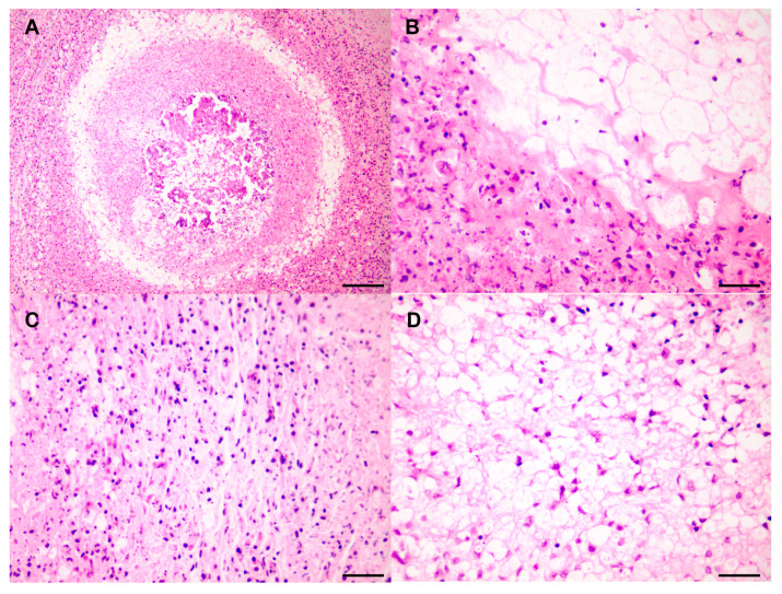
Figure 3.
Pathological observation of DDLPS. (A) The mass located on the mesentery, observed at low magnification. Scale bar = 120 µm. (B) Well–poorly differentiated transition areas. Scale bar = 30 µm. Immunohistochemistry results for (C) Poorly differentiated areas. Scale bar = 30 µm. (D) Well-differentiated areas. Scale bar = 30 µm.

