Figure 1.
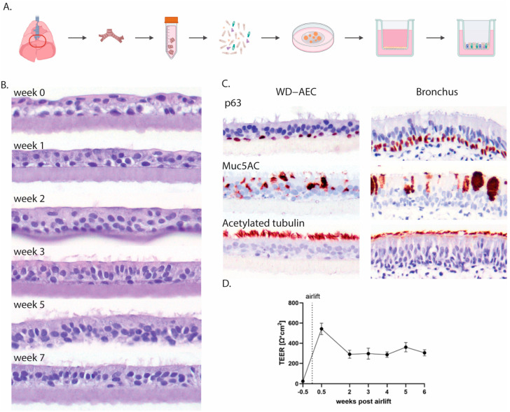
Establishment of AO-derived WD-AECs. (A) Schematic outline of the isolation of porcine bronchial epithelial cells from the primary bronchi which were subsequently grown as 3D organoids in an extracellular matrix before seeding in 2D on Transwell filters and culturing at air–liquid interface upon confluency. (B) Transverse histology sections over the course of 7 weeks showing the development of a pseudostratified ciliated respiratory epithelium. Hematoxylin and eosin stain, 40× objective. (C) Immunohistochemistry (IHC) staining of WD-AECs 7 weeks post-airlift and porcine bronchus epithelial cells. P63 staining to visualize basal cells, Muc5AC staining to visualize mucus (goblet cells) and acetylated tubulin staining to visualize cilia. WD-AEC resembles an in vivo bronchial epithelium in cellular composition and morphology, despite a reduced thickness. (D) Development of transepithelial electrical resistance (TEER) over the course of differentiation of WD-AECs. After an initial increase post-airlift, TEER values remained consistent. For B, C and D: Differentiation was followed for three separate donors. Representative data from one experiment are shown.

