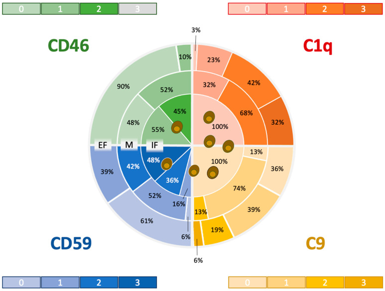
Figure 2.
Heatmap showing the distribution and expression levels of complement-activating factors C1q and C9 and complement-regulating factors CD46 and CD 59 in relation to FCoV-infected macrophages: Expression zones included the intrafocal area (IF), which colocalizes with FCoV-expressing macrophages; the marginal zone (M), which comprises the layer next to the circumference of FCoV-expressing cells; and the extrafocal zone (EF), which is separated from the IF by an immunonegative fringe. The color intensity mirrors the immunohistochemical scores (“the darker, the stronger”), whereas the percentages indicate the yield of lesions that expressed the markers at the respective strength per zone. Notably, high expression scores in the CRF were concentrated around FCoV-infected cells, whereas high expression scores were detected in the CAF in the outer zones only.

