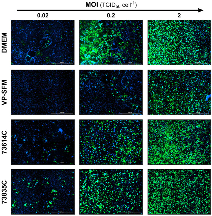
Figure 5.
Fluorescence images of MeV-infected static Vero cell cultures 3 dpi in SCM, SFM and CDMs at different MOIs. The expression of eGFP due to MeV infection is shown in green and cell nuclei are counterstained with DAPI (blue) in this merged image captured using the Cytation 3 (objective magnification: 2.5×, four (2 × 2) images per well, stitched).

