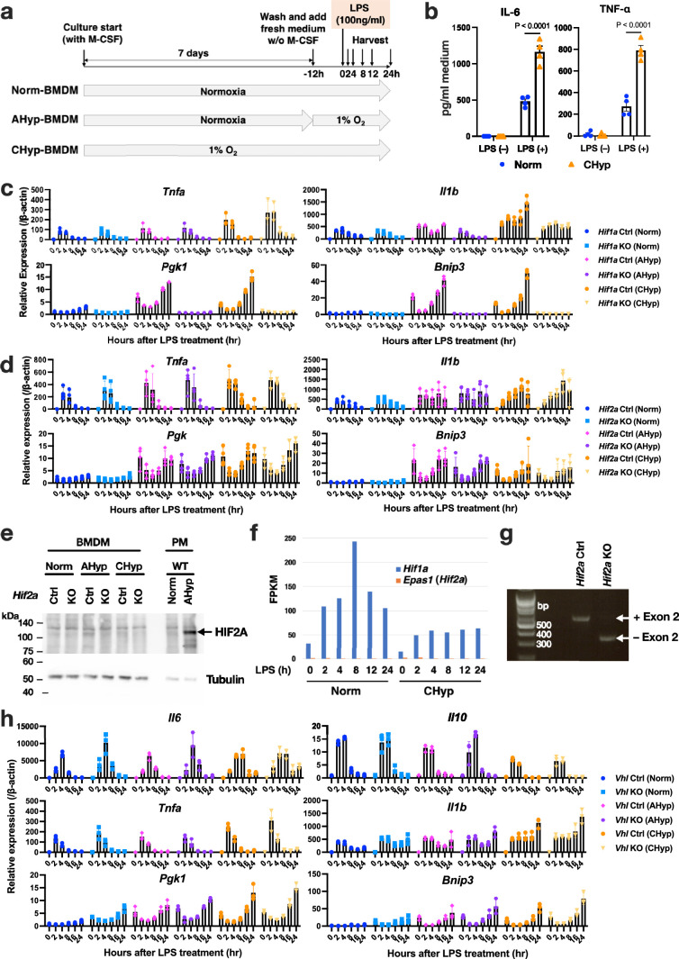
Extended Data Fig. 2. Inflammatory response of BMDMs differentiated under different oxygen tension.
a. Experimental design for BMDMs differentiation and stimulation with LPS (100 ng/ml). BMDMs were harvested for RNA-seq analysis at 0, 2, 4, 8, 12 and 24 h after LPS treatment. b. ELISA of cytokines in culture supernatant of BMDMs stimulated with or without LPS for 12 h (n = 4). Error bars represent S.E.M. Two-way ANOVA was conducted to evaluate statistical significance. c, d, h. Gene expression in BMDMs after LPS stimulation (n = 3). Norm, AHyp, and CHyp BMDMs generated from Hif1a mutant mice (c), Hif2a mutant mice (d), and Vhl mutant mice (h) were examined. Error bars represent S.E.M. e. Immunoblot analysis of Hif-2α protein. Tubulin was used as a loading control. Peritoneal macrophage (PM) of wild-type mice treated with acute hypoxia (AHyp) was examined as a positive control. A representative result from 2 independent experiments is shown. f. FPKM values of Hif1a and Epas1/Hif2a expression obtained from the RNA-seq analysis. g. PCR detection of exon 2 deletion of Hif2a gene in the genomic DNA of BMDMs after treatment with tamoxifen, performed once.

