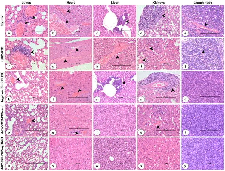
Figure 6.
Histopathological lesions in internal organs of mice at 14 days post challenge (dpc). Control (a–e), rNDV-R2B (f–j), Ingelvac CircoFLEX (k–o), rNDV-R2B-PCV (p–t), and rNDV-R2B-PCV-TMCT (u–y). (a) Lungs showed interstitial pneumonia with thickening of interalveolar spaces and marked congestion (arrow). H&E ×20. (b) Heart showed marked dilatation and congestion of myocardial blood vessels (arrow). H&E ×20. (c) Liver showed marked infiltration of inflammatory cells in portal triad (arrow). H&E ×20. (d) Kidneys showed marked infiltration of mononuclear cells (arrow). H&E ×20. (e) Lymph node showed congestion of blood vessels in cortex (arrow). H&E ×20. (f) Lungs showed moderate perivascular infiltration of lymphoid cells (arrow). H&E ×20. (g) Heart showed moderate to marked congestion of blood vessels (arrow). H&E ×20. (h) Liver showed marked congestion of blood vessels (arrow). H&E ×20. (i) Kidneys showed congestion and hemorrhages (arrow). H&E ×20. (j) Lymph node showed marked lymphoid depletion in cortex (arrow). H&E ×20. (k) Lungs showed mild to moderate thickening of interalveolar spaces and congestion (arrow). H&E ×20. (l) Heart showed mild to moderate congestion of myocardial vessels (arrow). H&E ×20. (m) Liver showed moderate infiltration of inflammatory cells in portal triad (arrow). H&E ×20. (n) Kidneys showed moderate infiltration of mononuclear cells (arrow). H&E ×20. (o) Lymph node showed mild lymphoid hyperplasia. H&E ×20. (p) Lungs showed mild congestion (arrow) and thickening of interalveolar spaces. H&E ×20. (q) Heart showed mild congestion of blood vessels (arrow). H&E ×20. (r) Liver showed mild congestion. H&E ×20. (s) Kidneys showed mild congestion (arrow). H&E ×20. (t) Lymph node showed moderate lymphoid hyperplasia. H&E ×20. (u) Lungs showed normal alveoli lined by flattened squamous type I pneumocytes. H&E ×20. (v) Myocardium showed normal branching cardiac fibers. H&E ×20. (w) Liver showed normal central vein with radiating cords of hepatocytes. H&E ×20. (x) Kidneys showed normal glomeruli and renal tubules in cortex. H&E ×20. (y) Lymph node showed marked lymphoid hyperplasia in cortex. H&E ×20.

