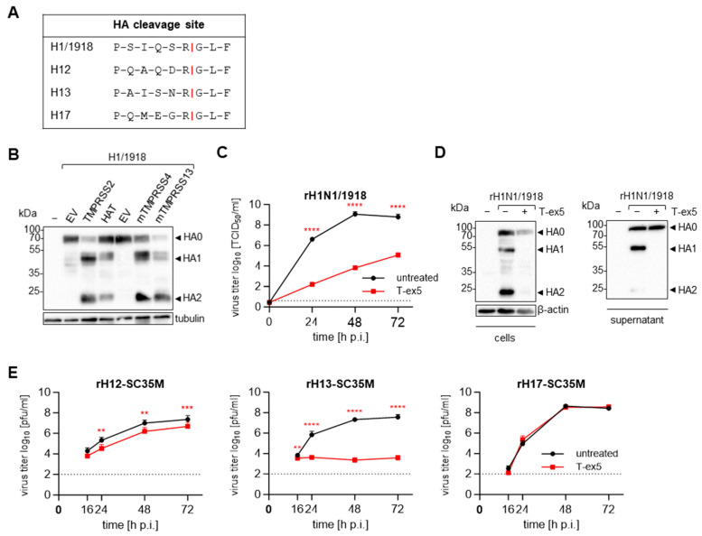
Figure 5.
TMPRSS2 dependency of H1N1/1918, H12, H13 and H17. (A) Sequence alignment of the cleavage sites from different HAs. (B) HEK293 cells transiently expressing the H1/1918 and human TMPRSS2 or HAT, or murine TMPRSS4 or TMPRSS13 were subjected to SDS-PAGE at 48 h p.t. Western blot analysis was performed with an antibody targeting H1. Tubulin was stained as a loading control. The result shown is representative of three independent experiments. EV = empty vector. (C) Calu-3 cells were treated with 25 µM T-ex5 for 24 h before being infected with rH1N1/1918 at an MOI of 0.001. Viral titers in the supernatant sampled at the indicated time points were determined via TCID50 endpoint titration. The data shown are the mean + SD of three individual experiments. The dotted lines indicate the LOD. (D) The cell lysates and supernatants (SN) of Calu-3 cells treated with 25 µM of T-ex5 for 24 h and then infected with rH1N1/1918 at an MOI of 1 for another 24 h were subjected to SDS-PAGE, and an antibody targeting H1 was used for the Western blot analysis. ß-actin was used as a control. The result is representative of three independent experiments. (E) Calu-3 cells pre-treated with 25 µM were infected with recombinant viruses containing avian H12 or H13 or bat-derived H17 and sharing the other seven gene segments of SC35M at a low MOI of 0.01–0.0001. The supernatant of infected cells was harvested at indicated time points and the viral titers were determined via an immune plaque assay. The experiments were performed in triplicate and the data shown are mean + SD. The dotted lines indicate the LOD. (C,E) Statistically significant differences in viral titers at each time point compared to untreated controls were determined by performing unpaired t-tests. ns = not significant, ** = p < 0.01, *** = p < 0.001, **** = p < 0.0001.

