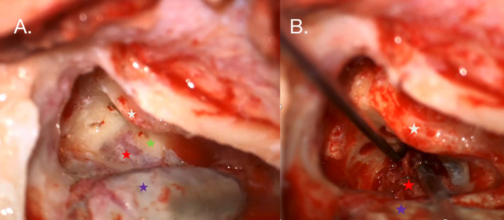
Figure 3.
(A) Intraoperative photography taken during the right ear procedure. An antromastoidectomy with posterior tympanotomy was performed. The facial nerve (white star) is visualized along the tympanic and mastoid segments. The white arrow indicates the tumor mass (red star) shining under the bone posterior to the posterior semicircular canal (green star), which is partially infiltrated by the tumor. The posterior semicircular and horizontal semicircular canals were drilled out to reveal the extent of the tumor. (B) The sigmoid sinus is skeletonized (violet star) and compressed by suction to visualize the highly vascularized tumor.

