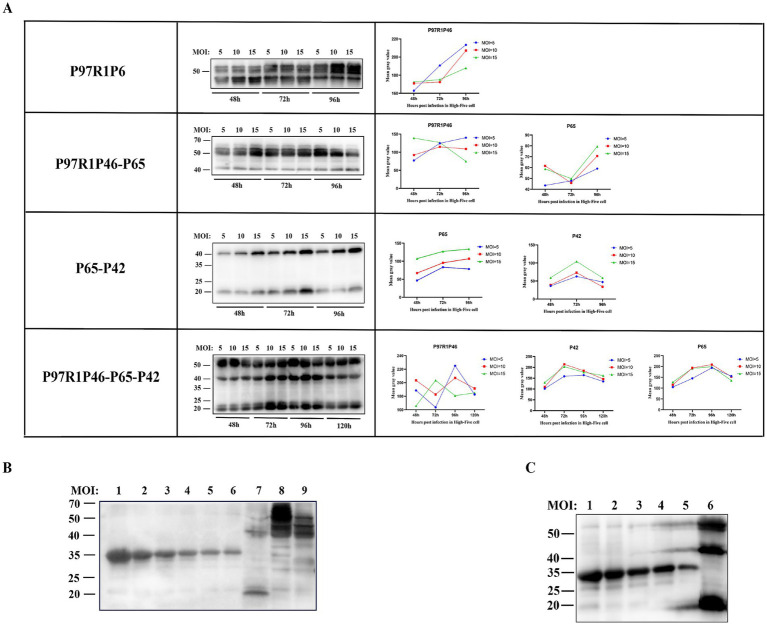
Figure 4.
Condition optimization and semi-quantitative analysis of recombinant proteins. (A) Western blot analysis of P97R1P46, P97R1P46-P65, P65-P42, and P97R1P46-P65-P42 proteins under different MOI and time conditions. (B) Semi-quantitative analysis of recombinant proteins. Lanes 1–6 are, in order, 800 μg/mL, 500 μg/mL, 250 μg/mL, 125 μg/mL, 62.5 μg/mL, and 31.25 μg/mL concentrations of standard protein; lane 7 is Ac-P65-P42 infected cell lysate; lane 8 is Ac-P97R1P46 infected cell lysate. Lane 9 is Ac-P97R1P46-P65 infected cell lysate. (C) Semi-quantitative analysis of recombinant proteins. Lanes 1–5 are, in order, 500 μg/mL, 250 μg/mL, 125 μg/mL, 62.5 μg/mL, and 31.25 μg/mL concentrations of standard protein; lane 6 is Ac-P97R1P46-P65-P42 infected cell lysate.

