Fig. 5.
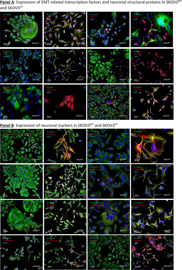
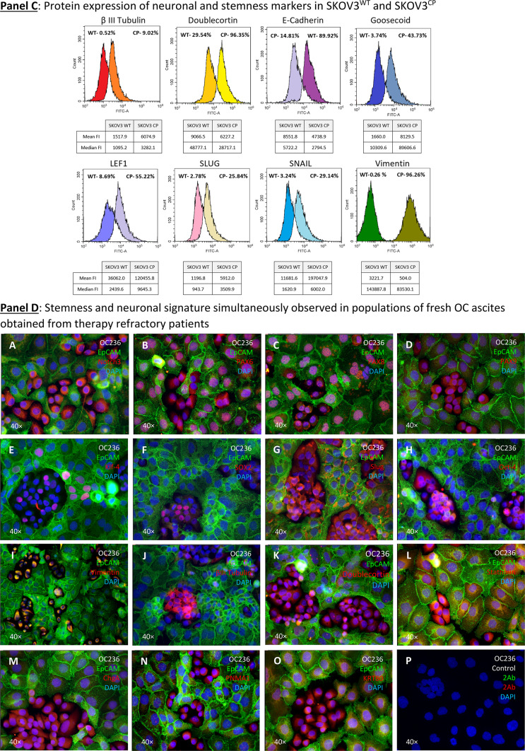
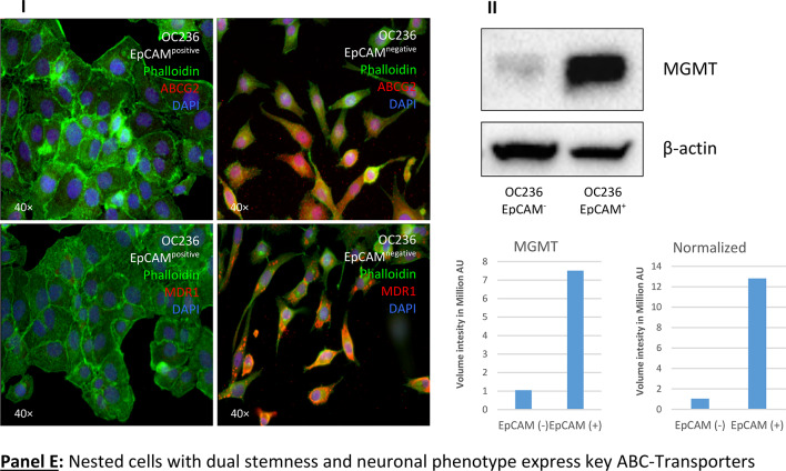
Expression of EMT-related transcription factors, neuronal structural proteins and MDR in SKOV3WT and SKOV3CP. Panel A: expression of cardinal transcription factors defining a neuronal phenotype and structural proteins. The basic helix-loop-helix (bHLH) HES1, the SRY (sex determining region Y) box 2 also known as SOX2, the paired box protein PAX6 and the basic helix-loop-helix transcription factor NeuroD, all well-known key players in neuronal development, are upregulated in SKOV3CP cells. Some of them like HES1, SOX2, and PAX6 show transcriptional upregulation in qPCR analysis. By the same token, structural proteins βIII-tubulin and Vimentin are upregulated, whereas the epithelial marker EpCAM is downregulated. Panel B: overexpression of important neuronal markers like MAP2, NF-L, N-cadherin, Tenascin C, Synaptophysin, Doublecortin and Stathmin in SKOV3CP. Importantly, the expression of these markers correlates with the neuronal morphology of the cells. Panel C: flow cytometric expression analysis of the EMT-related markers E-cadherin, Vimentin, Snail and Slug and the neuronal markers Doublecortin and Goosecoid. Panel D: stemness and neuronal signature in primary OC cells cultivated in 2D. Clusters of EpCAM negative cells with concurrent stemness and neuronal signature are observed. These nested cells are positive for different transcription factors (TFs, A-H) which are considered strong contributors to stem cell maintenance and cell differentiation. Of note, most TFs are mainly found in the cytoplasm of cells instead in the nucleoplasm, with the exception of PAX8, which is also localized to the nucleus (for reference, the RGB decomposition of pictures attached in the Supplemental Information). It is evident that those nested cells are not of epithelial nature by the lack of EpCAM and the expression of Vimentin (I), a structural protein typical for mesenchymal cells. Notably, those cells significantly express several neuronal markers like βIII-tubulin (J), a neuron-restricted microtubule protein, Doublecortin (K), Stathmin (L), the neuroendocrine marker Chromogranin A (ChgA) (M) and the dually neuron- and testis-specific protein PNMA1 (N). Interestingly, the type II intermediate filament Keratin 80 (KRT80) (O) was expressed in these nested cells, indicating that this filament is involved in early stages of tumor cell differentiation. P, secondary antibody control. Panel E: multidrug resistance in cells with stemness and neuronal phenotype. Nested cells isolated from primary cultures from ovarian carcinoma ascites express a multidrug resistance phenotype as judged by the expression of MDR1 and ABCG2. Cells were sorted into EpCAM positive and negative populations and analyzed by immunofluorescence. II: Expression of MGMT (Methylated-DNA–protein-cysteine methyltransferase) DNA repair enzyme in OC236, sorted by EpCAM expression. The EpCAM negative cells, which have a stemness/neuronal phenotype revealed no expression of MGMT whereas the EpCAM positive cells (epithelial) exposed an upregulation of this enzyme. This indicates that the EpCAM positive cells are more resistant to agents like cisplatin. ICC results from n = 6 experiments, FACS results from n = 3 experiments. Complete FACS histograms data can be found in Supplemental, Fig. 1.


