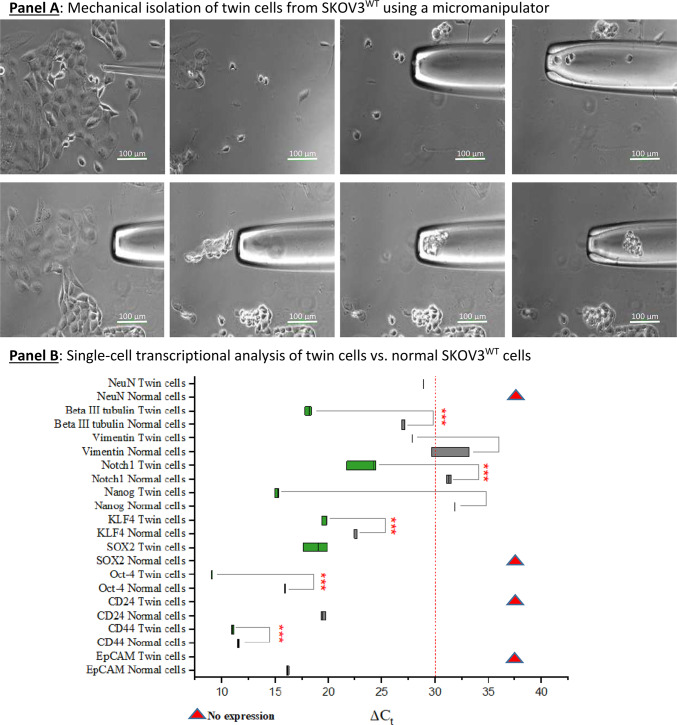
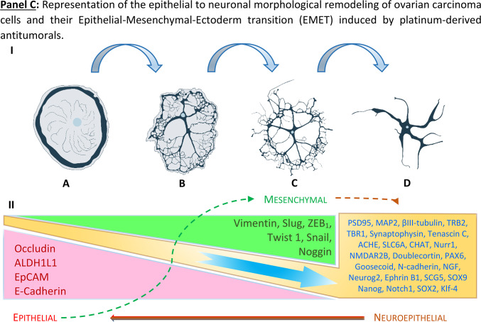
Fig. 6.
Isolation and analysis of “twin” cells present in SKOV3 cultures. Panel A: “twin” cells as shown in Fig. 3 (Panel A, A-D and Panel B, A-C) were subjected to mechanical isolation using a one-arm micromanipulator. At least 20 “twin” cells and a proportional amount of surrounding normal cells were collected for single-cell RNA isolation. Panel B: qPCR transcriptional analysis of “twin” cells vs. normal epithelia-shaped cells. A dual neuronal-stem signature is detected at the transcriptional level. Some markers were not detected (red triangles): NeuroN, SOX2 in normal cells and CD24 and EpCAM in “twin” cells. Panel C: I: Cartoon representing OC cells acquiring a neuron-like morphology under the influence of platinum-derived compounds as seen in Fig. 1. A large cell (A) gradually condenses its cytoplasm and starts forming dendritic tree-like structures (B & C). The cytoplasm is finally dismantled until fully formed dendrite-like structures are left (C & D). These morphological changes correlate with the expression of a plethora of neuronal-defining markers. II: Diagrammatic representation of the modulation of pathways involved in EMT and from this point to a “neuro-mesenchymal” inter-condition or EMET based on the transcriptional and protein expression profiles. SKOV3 cells have a basal expression of several neuronal markers, which is enhanced by exposure to sub-toxic concentrations of platinum-derived drugs. The expression of cardinal stemness markers like Nanog, Notch1, SOX2, and Klf-4 suggests the acquisition of cancer stem cells status. The cells that have moved to a neuroepithelial condition are in a transitory status of dormancy. Results are representative of n = 3 experiments.


