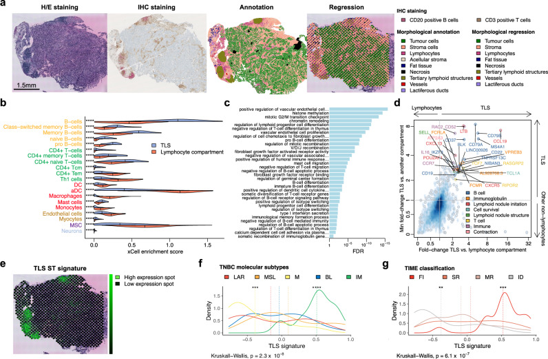
Fig. 5. Spatial characterization of tertiary lymphoid structures and development of a 30-gene TLS ST signature.
a Illustrative sample (ST_TNBC_ID 30) demonstrating TLS detection via CD3/CD20 IHC staining, along with morphological annotation of the H/E-stained ST slide (highlighted in khaki) and corresponding morphological regression (also in khaki). The same analyses were conducted across the entire cohort (except for IHC: N = 86), with regression performed on duplicates or triplicates of each ST sample. b Cell-type enrichment by xCell in TLS compared to the lymphocyte compartment in the ST TNBC cohort (N = 94). Median values are indicated by vertical lines. Only FDRs <0.05 are reported using two-sided Wilcoxon rank sum test. c Selected enriched biological pathways identified by GO: BP in TLS compared to the lymphocyte compartment in the ST TNBC cohort. Only FDRs <0.05 are reported using one-sided Wilcoxon rank sum test. d Scatter plot displaying 30 differentially expressed genes from the comparison of TLS with either lymphocyte (x-axis) or other non-lymphocyte compartments (y axis), composing the TLS ST signature in the ST TNBC cohort (N = 94). e Projection of the TLS ST signature expression (neon green) on the same TNBC sample (ST_TNBC_ID 30). f, g Distribution of TLS ST signature expression across TNBC molecular subtypes (N = 94) (f) and TIME classification (N = 93) (g) in the ST TNBC cohort. Dashed lines represent the mean signature by subgroup. Two-sided P values are from Kruskal–Wallis tests and Wilcoxon rank-sum tests (for comparisons of each class to all classes). FDRs were calculated using the Benjamini & Hochberg method to adjust P values (*FDR < 0.05 and ≥0.01; **FDR < 0.01 and ≥0.001; ***FDR < 0.001 and ≥0.0001; ****FDR < 0.0001). Source data and exact P values are provided as a Source Data file. aDC activated dendritic cells, BL basal-like, DC dendritic cells, FDR false-discovery rate, FI full inflamed, H/E hematoxylin and eosin, ID immune desert, IHC immunohistochemistry, IM immunomodulatory, LAR luminal androgen receptor, M mesenchymal, MR margin restricted, MSC mesenchymal stem cell, MSL mesenchymal stem-like, SR stroma restricted, Tcm central memory T cells, Tem effector memory T cells, Th1 type 1 helper, TIME Tumor Immune Microenvironment, TLS tertiary lymphoid structure.

