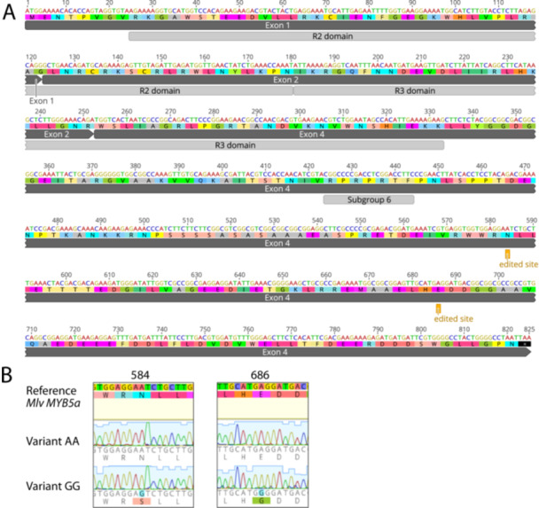
Figure 2.

Putative A‐to‐I editing sites in the exon 1‐2‐4 splice variant of M. l. variegatus MYB5a. (A) Dark gray bars show the exon structure from start codon to stop codon. Light gray bars show the DNA‐binding R2 and R3 domains common to all members of the R2R3 MYB gene family (Stracke, Werber, and Weisshaar 2001). “Subgroup 6” is a sequence motif that is conserved across all R2R3 MYB genes that encode activators of anthocyanin biosynthesis (Stracke, Werber, and Weisshaar 2001). The two putative A‐to‐I editing sites are each marked as “edited site.” (B) Chromatograms from MYB5a Variant AA and Variant GG. The two polymorphic sites are both located in the fourth exon of MYB5, 584 and 686 nucleotides downstream of the translation start site. Nucleotide and amino acid differences are highlighted. Sequences were obtained using Sanger sequencing and were visualized using Geneious R10. [Color figure can be viewed at wileyonlinelibrary.com]
