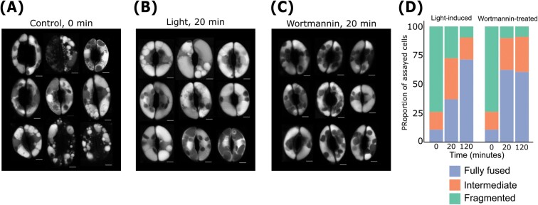
Figure 2.
Fusion due to wortmannin treatment proceeds more quickly than fusion due to a fusicoccin stimulus. (A) Dark-acclimated, ABA-treated Arabidopsis thaliana guard cells were imaged prior to treatment with a fusion-inducing stimulus. (B) Vacuole morphology after 20 min of fusicoccin and light treatment and (C) after 20 min of wortmannin treatment. (D) A qualitative survey of vacuole morphology reveals rapid wortmannin-driven fusion. We classified guard cells as having fragmented, fully fused or intermediate vacuole phenotypes at 0 min, 20 min, and 2 h after inducing fusion. The evolution of vacuole morphology was complete after 20 min in the wortmannin-treated cell group. Vacuole morphology in fusicoccin-treated cells continued to evolve after that time. Vacuoles were stained with BCECF. Chloroplasts (dark ovals inside guard cells) typically do not take up the vacuole stain. Source data for panel D available in Supplemental Table 1.

