
Fig. 1. Examples of pathological visualization of available AITL cases.
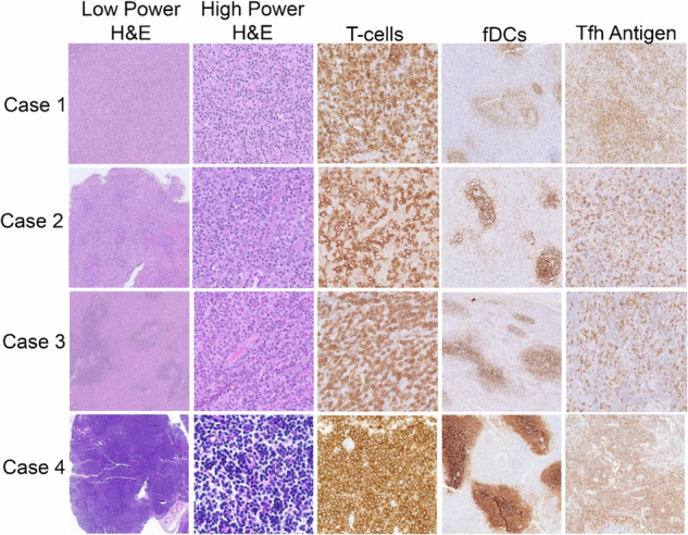
Low-power images (left column) of biopsied lymph nodes show varying degrees of architectural effacement by a paracortical infiltrate. High-power images (2nd to left column) of the paracortical infiltrate show atypical mature lymphocytes. T-cell-specific immunohistochemical stains (Case 1, BetaF1; Case 2, CD2; Cases 3 and 4, CD3) highlight the T-cell atypia. Follicular dendritic cell meshworks (fDCs) are focally mildly expanded in all cases (CD21 immunostain). T follicular helper cell (Tfh) antigens were expressed in all cases, illustrated by ICOS (Cases 1-3) and PD1 (Case 4) immunostains.