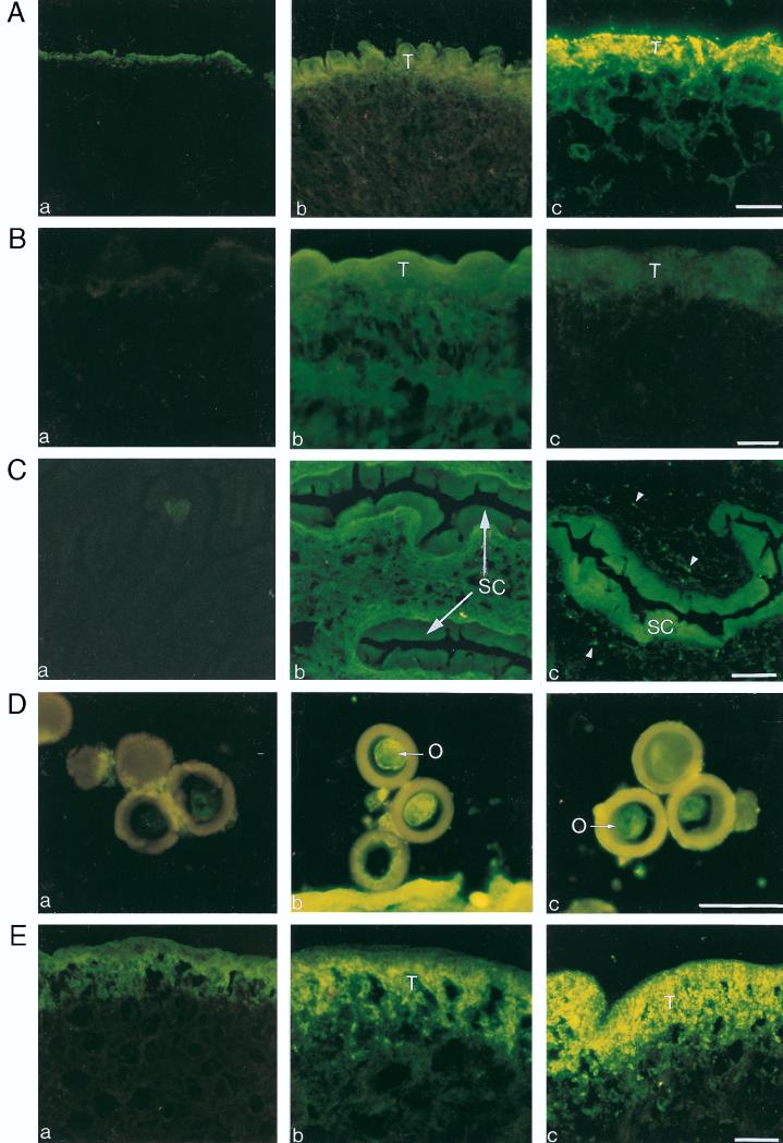
FIG. 1.
Immunofluorescence staining of T. crassiceps (A) and T. solium (B and C) cysticerci and of eggs (D) and adult tegument (E) of T. solium, incubated with sera from normal mice (a), 30-day T. crassiceps-infected mice (b), and pooled sera obtained 15 days after the booster of GK-1 (c). The labeled epitope is clearly evident in structures accessible to the immune system. It is intensively expressed in the tegument (T) of T. crassiceps cysticerci (A, panel c) and weakly in the T. solium cysticerci (B, panel c). It is strongly expressed in the cuticular folds of the spiral canal (SC) (C, panel c), in the oncosphere (O) (D, panel c), and in the distal tegument (T) of the tapeworm (E, panel c). The arrowheads (C, panel c) indicate the protonehridia. Bars = 40 μm.

