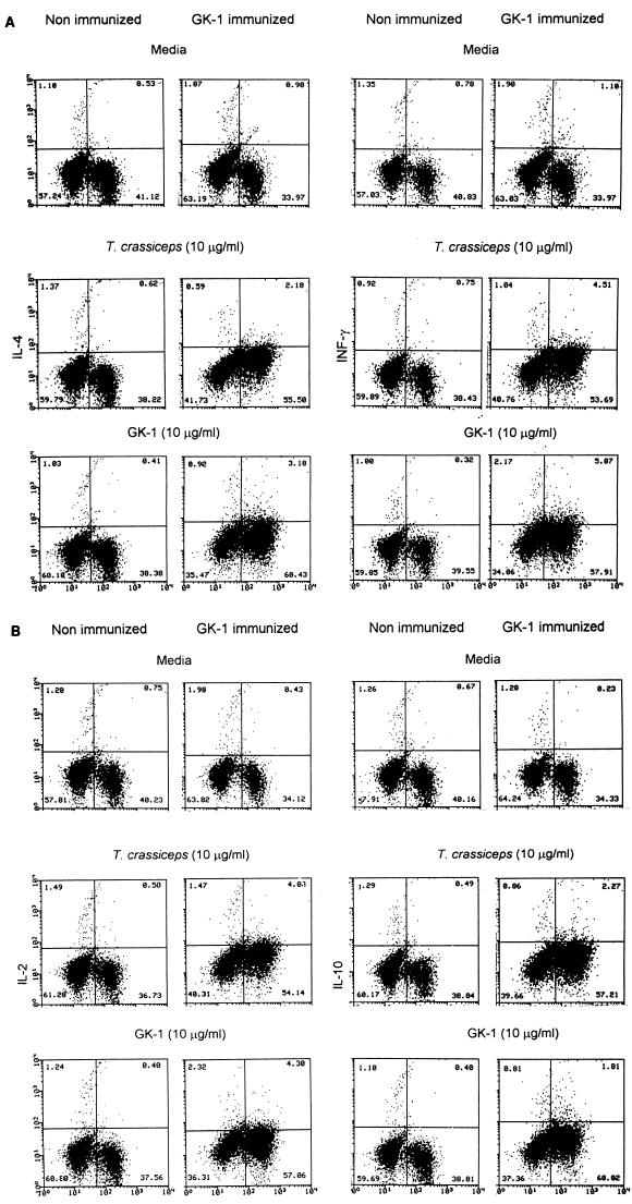
FIG. 3.
Spleen lymphocytes from nonimmunized (injected with saponin alone) and GK-1-immunized mice 60 h poststimulation were analyzed for intracellular cytokines (IFN-γ and IL-4 [A] and IL-2 and IL-10 [B]) and surface CD3+ staining by FACS. Cells had been dually stained with FITC (abscissa) and PE (ordinate). The percentage of cells in each quadrant of the dot plot is indicated. The data are representative of three experiments using different mice.

