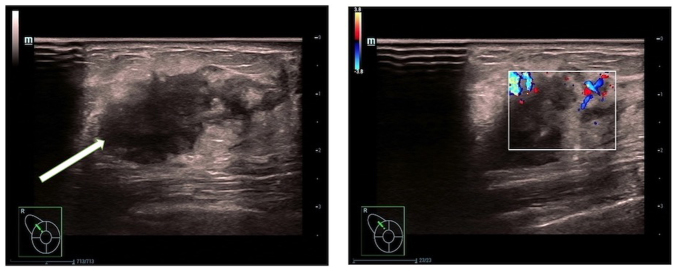
Figure 2.
Ultrasound images of the breast lesion. A low-echo nodule measuring ~22.1×14.4 mm is observed in the right breast, at the 11 o'clock position, approximately one finger-width away from the nipple and 5 mm from the skin surface (left panel). The nodule has an irregular shape and unclear boundary. Color Doppler flow imaging shows linear blood-flow signals within the nodule (right panel). These ultrasound findings suggest a solid nodule in the right breast, falling under Breast Imaging-Reporting and Data System classification 4a. A biopsy was recommended.

