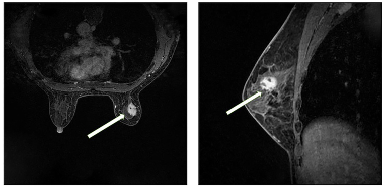
Figure 3.
Magnetic resonance imaging of the right breast. A circular shape with a slightly longer T2 signal is evident in the upper outer quadrant of the right breast, which. exhibits high signal intensity on diffusion-weighted imaging (left panel). The lesion is ~18.5×14 mm in size, and has irregular margins with visible lobulations and spicules. Contrast-enhanced doubly deprotonated-diethylenetriamine penta-acetic acid scanning (right panel) shows marked heterogeneous enhancement of the lesion, and the dynamic curve displays a plateau shape. The arrows indicate the lesion. Based on this imaging, the diagnosis was a lesion in the upper outer quadrant of the right breast, classified as Breast Imaging-Reporting and Data System 4b.

