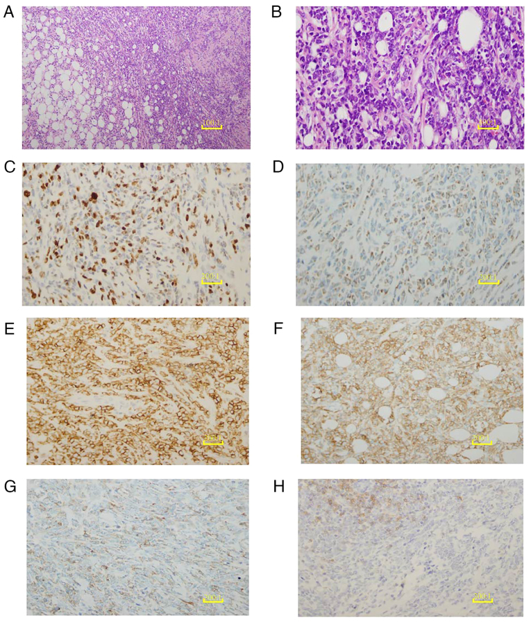
Figure 5.
Histopathological and immunohistochemical analysis of the breast mass. (A) H&E staining of the breast mass biopsy tissue reveals diffuse, sheet-like growth with disrupted ductal structures (scale bar, 10 µm). (B) The H&E-stained breast mass biopsy tissue is extensively infiltrated by medium-sized malignant cells with round follicular nuclei containing finely dispersed chromatin and small nucleoli (scale bar, 2.5 µm). (C) Ki-67 shows a proliferation index of 50%, serving as a marker for cancer cells. (D) Myeloid differentiation of the tumor cells is illustrated with a myeloperoxidase immunohistochemical stain. (E) Positive expression of CD43 is revealed by immunohistochemical staining, supporting the diagnosis of myeloid sarcoma. (F) Tumor cells surrounding the breast ductal epithelium are positive for CD117 immunohistochemical staining, indicating their myeloid origin. (G) The hematopoietic progenitor origin of the tumor cells is indicated by weakly positive CD34 immunohistochemical staining. (H) Weak CD19 immunohistochemical staining indicates the reduced or abnormal expression of B-cell markers in the tumor cells. (C-H), scale bar, 5 µm. H&E, hematoxylin and eosin; CD, cluster of differentiation.

