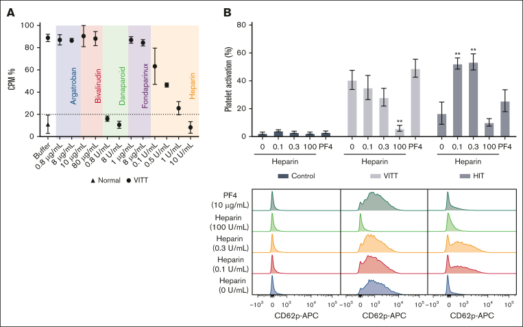
Figure 2.
Danaparoid and UF heparin block platelet activation by VITT IgG. (A) SRA shows that VITT-induced platelet activation is inhibited in the presence of therapeutic (0.8 μg/mL) and high-dose danaparoid (8 μg/mL) and high-dose UF heparin (10 U/mL) but not argatroban (0.8 and 8 μg/mL), bivalirudin (10 and 80 μg/mL), fondaparinux (1 and 8 μg/mL), or low-dose UF heparin (0.1, 0.5, and 1 U/mL). Data are shown as the mean ± standard deviation (SD). The data are representative of 3 patient samples with VITT. (B) IgG-induced platelet activation in the absence or presence of heparin (0.1, 0.3, and 100 U/mL) or PF4 (10 μg/mL). The platelets were treated with control (normal, n = 4), VITT (n = 6), or HIT IgG (n = 3). Data are shown as the mean ± standard error of the mean. ∗∗P < .01, relative to VITT or HIT IgG without heparin or PF4. Representative flow cytometry histogram plots showing platelet activation (anti-CD62p APC) following treatment with control, VITT or HIT IgG in the absence or presence of heparin or PF4. APC, allophycocyanin; CPM, counts per minute.

