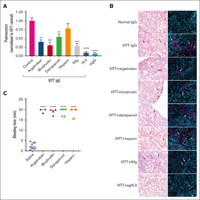
Figure 4.
Effect of nonheparin anticoagulants, heparin, IVIg, and IV.3 in VITT mice. (A) Mouse lungs from FcγRIIa+/hPF4+ mice treated with VITT IgG, with or without anticoagulants, were extracted and analyzed for thrombosis by fluorescence using the IVIS SpectrumCT. (B) Representative images of hematoxylin and eosin-stained and fluorescence-stained lung sections. Images were acquired with a 10× objective using an inverted Olympus CKX53 microscope. Platelets (anti-CD42c) and DNA (4′,6-diamidino-2-phenylindole [DAPI]) are shown as magenta and cyan, respectively. Arrows indicate clots. Scale bar, 50 μm. (C) Mice treated with argatroban, bivalirudin, danaparoid or UF heparin at therapeutic concentrations had prolonged bleeding times compared with the saline control. The bleeding time was measured over 20 minutes. Data are shown as mean ± SD. ∗P < .05; ∗∗P < .01; ∗∗∗P < .001; ∗∗∗∗P < .0001, relative to VITT IgG (A) or saline (C).

