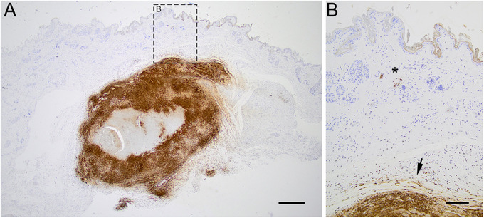
FIGURE 4.

Microphotograph of the immunohistochemistry visualizing the bacteria Francisella tularensis (brown stain) in the skin at the location of a tick bite in a F. tularensis subsp.‐holarctica positive hare (ID A). (A) Section of skin at the site of the tick bite. F. tularensis visualized in lytic lesion in the subcutis, bar = 500 µm. (B) Close‐up of (A), bacteria visualized in the cytoplasm of vascular endothelial cell (asterisk) and lytic lesion in the subcutis (arrow), bar = 200 µm.
