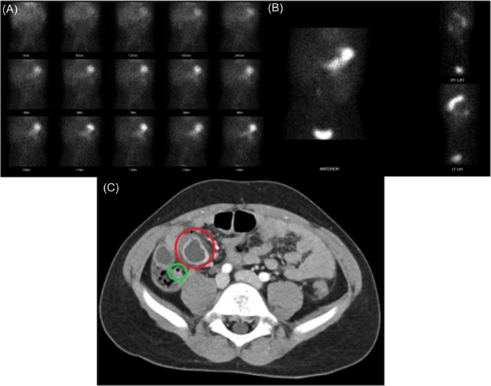
Figure 2.

Imaging findings of two patients with Meckel's diverticulum. (A, B) The first 15 min (A) and 1‐h images (B) of a Meckel's scan in a 5‐year‐old boy with abdominal pain, anemia, and bloody stools. No preimaging medication was received. Physiologic radiotracer uptake is seen, and no abnormal radiotracer accumulation is identified. (C) CT abdomen and pelvis with IV contrast in a 7‐year‐old boy with abdominal pain who previously had a negative Meckel's scan and negative laparoscopy. A fluid collection with thick enhancing wall, inflammatory fat stranding, and a punctate focus of extraluminal air along its anterior margin (red circle) suggested perforation. A normal appendix was identified (green circle). CT, computed tomography.
