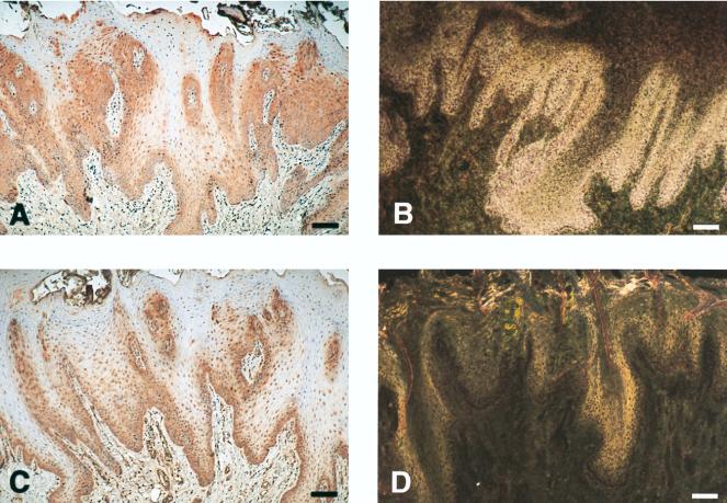
FIG. 2.
hCAP18 is expressed in the squamous epithelium of the tongue and colocalizes with immunoreactivity for IL-6. (A) Section of tongue demonstrating positive immunostaining for hCAP18 in the epithelium. Immunoreactivity (red precipitate) is most pronounced in the basal cell layers, but scattered immunopositive cells can also be seen in the suprabasal layers. (B) Section from the same tissue hybridized with antisense 35S-labeled cRNA probe for hCAP18 mRNA. Intense autoradiographic signal for hCAP18 mRNA (appears as white grains under dark-field illumination) is seen in the lower portion of tongue epithelium. (C) Immunoreactivity for IL-6 is seen in tongue epithelium with the same pattern as for hCAP18 and in submucosal blood vessels. (D) Hybridization with the control sense probe for hCAP18 lacks the autoradiographic signal for hCAP18 mRNA. The shiny appearance is due to the autofluorescence of the tissue. Bars, 100 μm.

