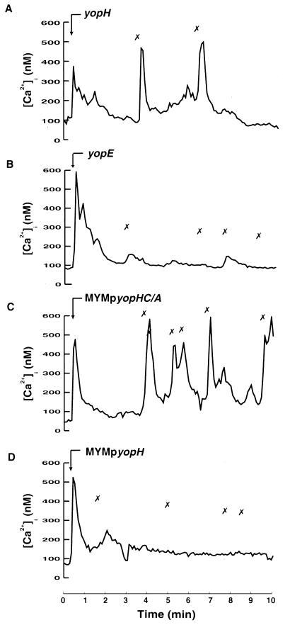
FIG. 4.
Involvement of YopH in inhibition of intracellular Ca2+ signaling. Time courses of [Ca2+]i in single neutrophils during attachment of different Y. pseudotuberculosis strains are shown. Adherent neutrophils were exposed to the Yersinia yopH mutant (A), yopE mutant (B), MYM transcomplemented with yopHC/A (C), or MYM transcomplemented with wild-type yopH (D) at a calculated bacterium/cell ratio of 50:1 (arrow). The “✗” symbols indicate contact between the bacteria and the neutrophil. Attachment of bacteria was visually observed on a video screen, and this was correlated with the [Ca2+]i transients. Representative time courses are presented (of 9 to 12 separate experiments). The mean numbers of [Ca2+]i transients are shown in Table 2.

