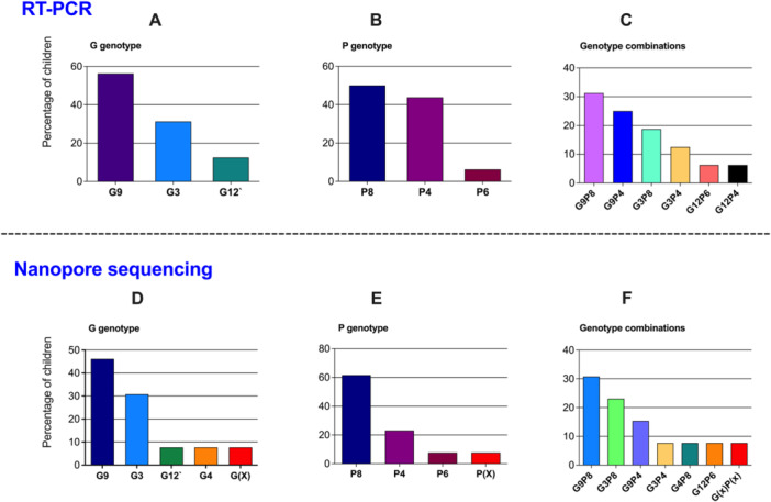
Figure 2.

Genotypes identified by RT‐PCR and nanopore sequencing from the same samples. Upper panel: (A) G (VP7) typing, (B) P (VP4) typing, and (C) genotypes combination of the rotavirus strains typed by RT‐PCR (n = 16, two samples were excluded because of poor RNA quality). Lower panel: (D) G (VP7) typing, (E) P (VP7) typing, and (F) genotypes combination by nanopore sequencing (n = 13, three samples were excluded because of poor RNA quality). The graph presents percentage of rotavirus strains of G and P genotypes detected in the children; (X: nontypeable).
