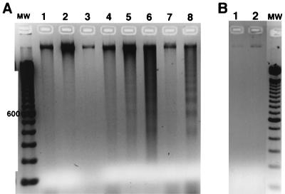
FIG. 4.
Internucleosomal breakdown of host cell DNA after EPEC infection. T84 cells were treated with inhibitors or infected with E. coli for 6 h; medium was replenished once at 2 h, at which time wortmannin was readded. Cells were recovered in 70% ethanol and subjected to extraction with phosphate-citric acid as described in Materials and Methods and then analyzed by agarose gel electrophoresis. A negative image of the ethidium bromide-stained gel is shown. (A) Extracts from human cells; (B) extracts from E. coli alone; MW, 100-bp DNA molecular size markers, with the 600-bp marker indicated. (A) Lane 1, uninfected, control T84 cells; lane 2, cells treated with 100 nM wortmannin alone; lane 3, cells treated with 200 μM genistein alone; lane 4, cells infected with EPEC strain E2348; lane 5, cells treated with EPEC plus wortmannin; lane 6, cells treated with E2348 plus genistein; lane 7, cells infected with strain HS alone; lane 8, H9 leukemia cells treated with doxorubicin (2 μg/ml) for 16 h as a positive control. (B) DNA extracts from E. coli HS (lane 1) and EPEC strain E2348 (lane 2). In panel B, the number of bacteria subjected to the extraction was about five times greater than that calculated to be present in panel A.

