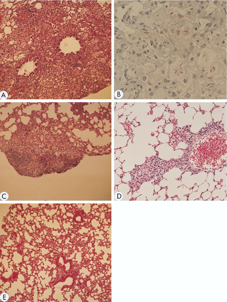
FIG. 3.
Histologic examination of lung tissues. Mice were killed 7 weeks after infection, and formalin-fixed sections were stained with hematoxylin and eosin (A, C, D, and E) and for acid-fast bacilli (B). (A) Tissue from IL-18-KO mice infected with the Kurono strain. A large, indiscrete granuloma with foamy macrophages is noted. Magnification, ×100. (B) Tissue from IL-18-KO mice infected with the Kurono strain. Mycobacteria stained red and are recognized in the granuloma by Ziehl-Neelsen staining. Magnification, ×600. (C) Tissue from WT mice infected with the Kurono strain. A small, discrete granuloma was formed. Magnification, ×100. (D) Tissue from IL-18-KO mice infected with the Kurono strain and treated four times subcutaneously with recombinant IL-18. The granuloma became smaller. Magnification, ×200. (E) Tissue from WT mice infected with BCG Pasteur. No granuloma was recognized. Alveolar septal thickening was noted. Magnification, ×100.

