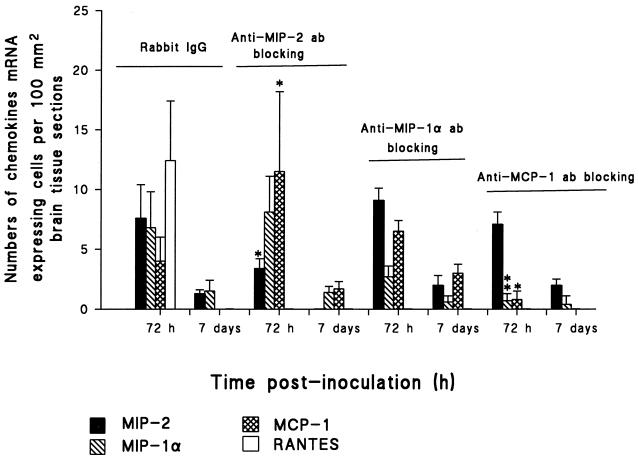
FIG. 6.
Effect of MIP-2, MIP-1α, and MCP-1 Abs on chemokine mRNA expression. Four groups of six infant rats received either anti-MIP-2, anti-MIP-1α, and anti-MCP-1 Ab (5 μg/day) or isotype control Ab (5 μg/day) i.p. 30 min before Hib inoculation. Rats then received i.p. injections with 100 μl containing 105 CFU of Hib per ml. Results are given as mean numbers of cells expressing mRNA for MIP-2, MIP-1α, MCP-1, and RANTES per 100 mm2 of surface area, detected by in situ hybridization as described in Materials and Methods (error bars, standard deviations). Brain sections were obtained from anti-MIP-2-treated and isotype control-inoculated groups at 72 h and 7 days p.i. For each panel, the significance of differences between treated and untreated groups of Hib-inoculated rats was determined by Student’s t test. ∗∗∗, P < 0.001; ∗∗, P < 0.01; ∗, P < 0.05.

