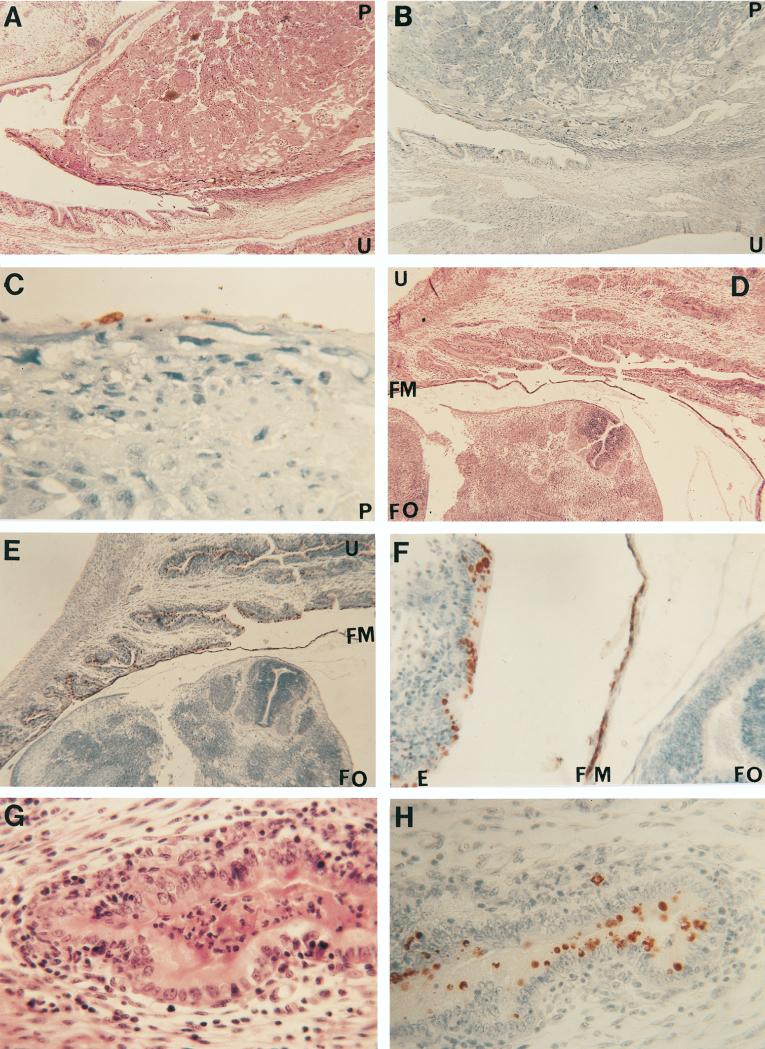
FIG. 1.
(A to C) Histological section at the placental site of insertion stained with H&E (A [magnification, ×30]) and an IHC stain for C. trachomatis (B [×30] and C [×250]). The overall architecture of the placenta and the uterine wall is well preserved (A). Chlamydial inclusions can be detected in the endometrium (B) and in the periplacental bilaminar omphalopleure (B and C). (D to F) Section of the fetus and uterine wall stained with H&E (D [×30]) and an IHC stain for C. trachomatis (E [×30] and F [×160]). Fetal tissues appear normal and at a developmental stage corresponding to 14 to 15 days of gestation (D). Chlamydial inclusions can be observed in the endometrium and in splanchnopleure of the yolk sac (E and F). (G and H) Uterus stained with H&E (G [×250]) and the IHC stain for C. trachomatis (H [×250]). A moderate acute and chronic inflammatory reaction (G) and multiple chlamydial inclusions (H) can be observed in the endometrium. Abbreviations: E, endometrium; FM, fetal membranes; FO, fetal organs; P, placenta; U, uterus.

