Figure 1.

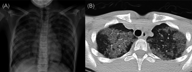
Pretreatment images (A) chest X‐ray; bilateral ground‐glass opacity, batwing pattern. (B) The chest computed tomography; crazy paving pattern, thickening of secondary lobules, and septal thickening.

Pretreatment images (A) chest X‐ray; bilateral ground‐glass opacity, batwing pattern. (B) The chest computed tomography; crazy paving pattern, thickening of secondary lobules, and septal thickening.