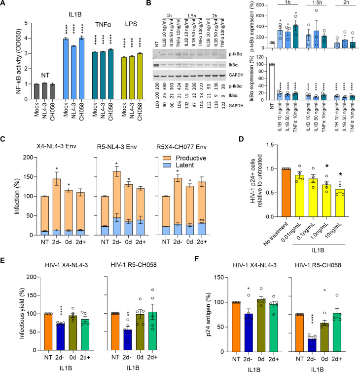
Figure 5. In vitro IL1B activates NF-κB, increases HIV proviral transcription, and inhibits spreading infection.
A) Effects of IL1B on NF-κB activity were assessed using A549 NF-κB reporter cells. Cultures were treated with IL1B, LPS, and TNFα and infected with VSV-pseudotyped NL4-3, CH058, or Mock control. After 24h the Alkaline Phosphatase Blue Microwell assay was performed with OD650 values relative to no treatment control (NT) reflecting NF-κB expression which is shown on the Y-axis. B) PBMC from 3 donors were treated with IL1B or TNFα and examined for IκBα phosphorylation as described in the methods section. Graphs present the protein expression from these donors; unpaired t test, *p<0.05, ****p<0.0001. C) Effects of IL1B in vitro when HIV was quantified after a single round of infection. Plots show the relative proportions of pMorpheus-V5 latently (blue) or productively (orange) infected PBMC in cultures treated with IL1B prior to, simultaneously, or after transduction with Env viral particles carrying the indicated Env protein. The data represent the average of 3 individual healthy donors, with error bars representing the average ±SEM, and statistical significance was established using unpaired t tests; *p<0.05, **p<0.002. D) Effects of IL1B on spreading HIV-1 infection in cell culture. Using HIV-1 YU-2, bar plots display the relative p24-positive cell fractions after pre-treatment with increasing concentrations of IL1B (from 0.01–10.0 ng/mL, 10-fold increments) across four different donors. E, F) Bar plots display the average infectious virus yields (E) and p24 antigen levels (F) at 4 days post-infection relative to the no IL1B treatment controls normalized to 100%; unpaired t test, *p<0.05, **p<0.002, ***p<0.0002. Corresponding replication curves are shown in Fig. S8.

