Figure 3.
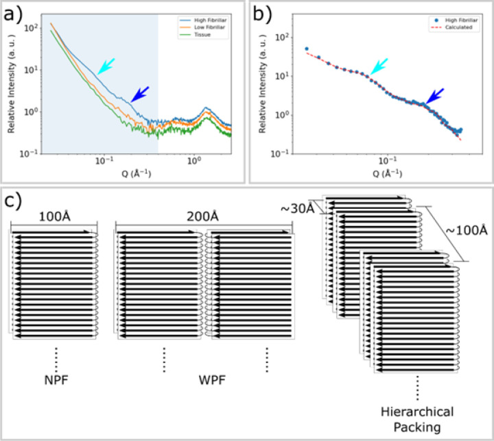
SAXS from fibrillar tau exhibits distinctive peaks. a) The SAXS profiles from tissue devoid of tau aggregates (green), containing low fibril content (orange) and having high levels of fibrillar tau (blue) as identified by the intensity of the pronounced WAXS reflection at 4.7 Å spacing. Patterns exhibiting strong cross-β related features in the WAXS regime (Q ~ 1.36 Å−1) also exhibit distinctive features in the SAXS regime, highlighted by cyan and blue arrows at Q ~ 0.07 Å−1 and ~ 0.2 Å−1. b) Background-subtracted SAXS intensities correspond well with those calculated from a hierarchical model of fibrillar organization exhibiting hierarchical packing with limited variation in fibril-fibril distances. c) A diagram of the narrow pick filament (NPF), the wide pick filament (WPF) and the model of hierarchical packing used to fit the observed data. Inter-fibrillar distances of 30 Å and 100 Å are representative of the polymorphic hierarchical organization indicated by the shape of the SAXS scattering as detailed in Figure S4.

