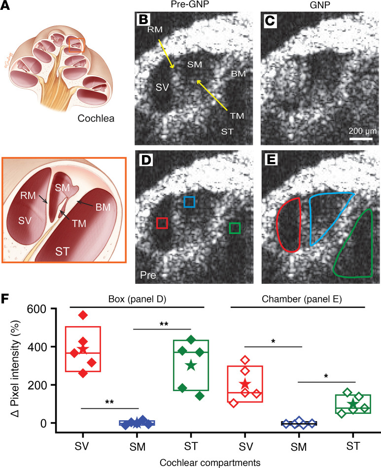
Figure 6. In vivo real-time monitoring of gold nanoparticles (GNPs) injected through the PSCC.
(A) The top presents the cross-sectional image of the cochlea and the bottom shows the region outlined in orange rotated to match the OCT images. Cochlear chambers are labeled: scala vestibuli (SV), scala media (SM), and scala tympani (ST). These chambers are separated by Reissner’s membrane (RM) and basilar membrane (BM). Tectorial membrane (TM) is within the SM compartment. Optical coherence tomography (OCT) images with the same labeling as panel A show (B) before GNP (50 nm) injection and (C) after GNP injection. (D) Boxes show small regions of interest (ROIs) that avoid the organ of Corti tissue structure. (E) Entire chamber labeling includes the organ of Corti structures. Scale bar: 200 μm (B–E). (F) The change in fluorescence intensity with GNP injection. Closed symbols are box ROIs from panel D and open symbols are the entire chamber ROIs from panel E. Boxes, SD; stars, mean; lines, median. *P < 0.05; **P < 0.01 by paired, 2-tailed Student’s t test. Two-way ANOVA with Tukey’s post hoc test also revealed significant differences between the SM and SV, as well as between the SM and ST, in both the box and chamber regions.

