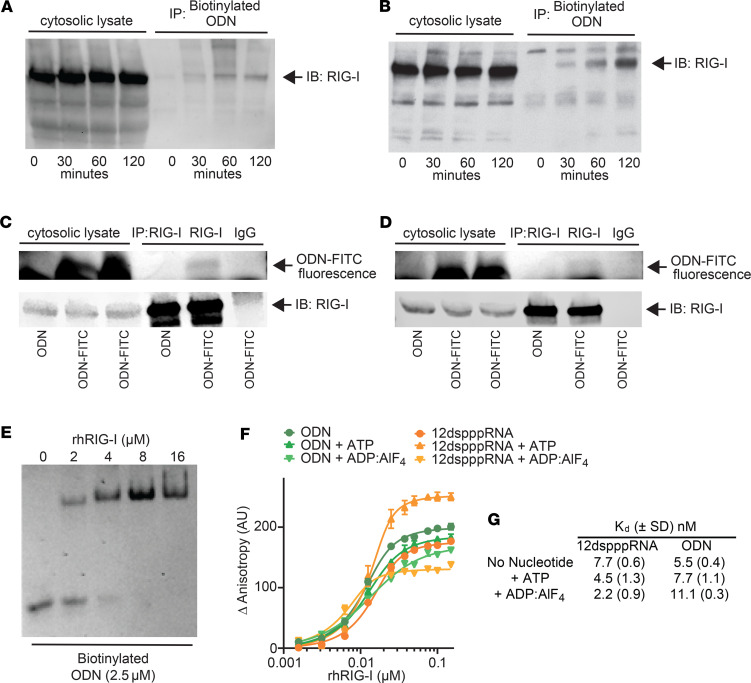
Figure 2. RIG-I is a cytosolic receptor for ODN.
(A and B) Cytosolic lysates from biotinylated-ODN-treated (A) human HBEC3-KT or (B) murine MLE-15 lung epithelial cells were streptavidin precipitated and probed with anti–RIG-I antibody. (C and D) Cytosolic lysates from FITC-labeled ODN-treated (C) HBEC3-KT or (D) MLE-15 cells were immunoprecipitated with anti–RIG-I antibody or IgG isotype control. Fluorescence from FITC-labeled ODN was detected by LI-COR imaging. RIG-I was probed with anti–RIG-I antibody. (E) Electrophoresis mobility shift assay of purified rhRIG-I protein incubated with ODN. (F) Fluorescence polarization anisotropy of rhRIG-I exposed to ODN or RNA in the presence or absence or ATP or ADP-AlF4. (G) The equilibrium binding constant (Kd) values of the corresponding fluorescence anisotropy binding curves are shown in F. rhRIG-I, recombinant human RIG-I; ADP-AlF4, adenosine diphosphate–aluminum fluoride. Data are shown as mean ± SD.

