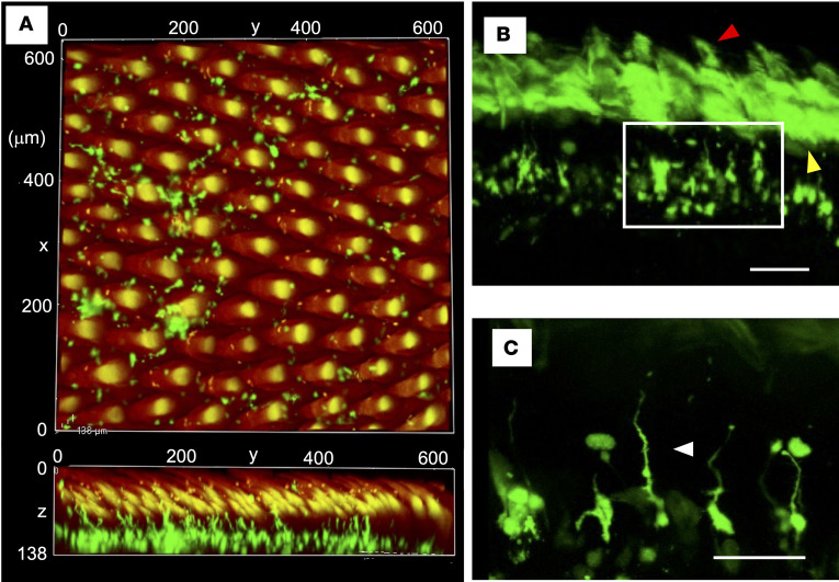
Figure 2. DCs in the dorsal tongue.
Images of the dorsal tongue acquired using confocal laser microscopy. (A) The posterior dorsal surface of the tissue-cleared tongue was observed. Upper: vertical image. Lower: sagittal image (Supplemental Video 1). (B) The surface of the coronal section of the tissue-cleared tongue was observed. Yellow and red arrowheads in B point to the autofluorescence of epithelium and filiform papillae, respectively. (C) Enlarged image of the area enclosed by the white square in B. Green, CD11c-YFP. Red, autofluorescence. The arrowhead points to the elongated dendrite to the cryptic bottoms on the dorsal surface. Scale bars: 100 μm (B) and 50 μm (C).

