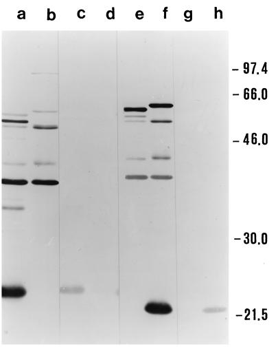
FIG. 3.
Western blot analysis of B. anserina strains. Lanes: a, c, e, and g, strain Es; b, d, f, and h, pathogenic strain Ni-NL. Lanes were probed as follows: a and b with MIAF to Es, c and d with Ab-SEE to Es, e and f with MIAF to Ni-NL, and g and h with Ab-SEE to Ni-NL. Molecular sizes (kilodaltons) are shown on the right.

