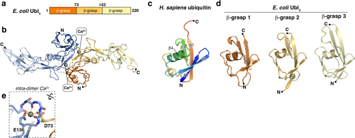
Figure 2. Crystal structure of E. coli Ubl3.
(a) Domain structure of E. coli Ubl3. (b) Crystal structure of an E. coli Ubl3 dimer, with one protomer colored dark orange/light orange/light yellow and the second protomer colored dark blue/medium blue/light blue. Bound Ca2+ ions are shown as gray spheres. (c) Structure of H. sapiens ubiquitin (PDB ID 1UBQ) 36, colored as a rainbow from N-terminus (blue) to C-terminus (red) and with β-strands 1–5 labeled. (d) Structures of E. coli Ubl3 β-grasp domains 1–3, colored as in panel (b) and aligned with the structure of H. sapiens ubiquitin. (e) Close-up of the intra-dimer Ca2+ ion bound by E. coli Ubl3 indicated by a dotted box in panel (b). See Figure S3a for a sequence alignment of Ubl3 β-grasp domains 1–2 showing conservation of this site.

