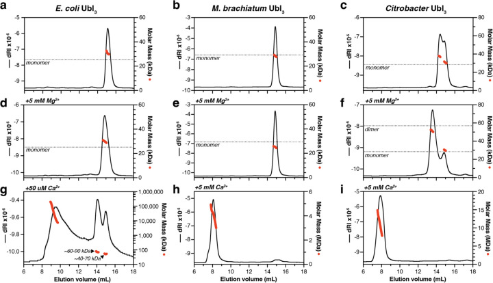
Figure 4. Ubl3 proteins oligomerize in the presence of Ca2+.
(a-c) SEC-MALS analysis of E. coli Ubl3 (a), M. brachiatum Ubl3 (b), and Citrobacter Ubl3 (d) in the absence of divalent cations. Black lines indicate dRI (change in refractive index; left Y axis) and red dots indicate measured molar mass (right Y axis; shown in logarithmic scale). The expected molar mass of a monomer species is shown as a dotted line. (d-f) SEC-MALS analysis of E. coli Ubl3 (d), M. brachiatum Ubl3 (e), and Citrobacter Ubl3 (f) in the presence of 5 mM MgCl2. The expected molar mass of monomer and dimer species are shown as dotted lines. (g-i) SEC-MALS analysis of E. coli Ubl3 in the presence of 50 µM CaCl2 (g), M. brachiatum Ubl3 in the presence of 5 mM CaCl2 (h), and Citrobacter Ubl3 in the presence of 5 mM CaCl2 (i).

