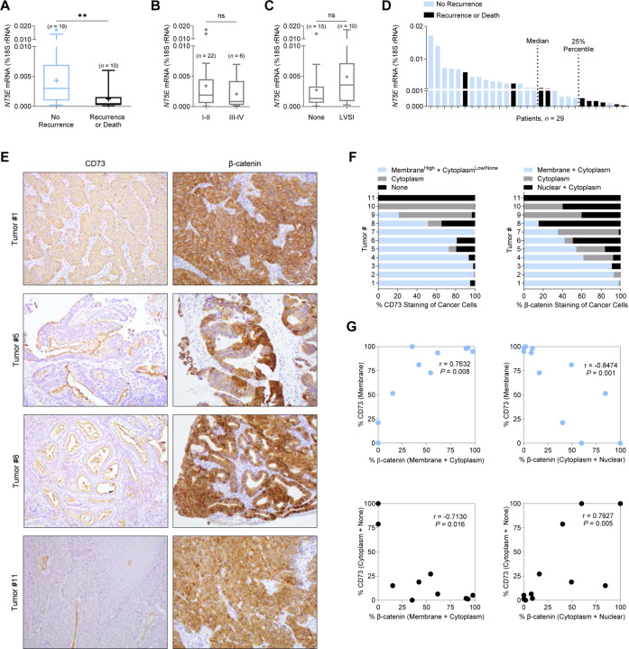
Figure 1. Loss of CD73 associates with poor patient outcomes and increased cytoplasmic/nuclear β-catenin in endometrial tumors with exon 3 CTNNB1 mutations.
(A-D) NT5E mRNA expression in exon 3 CTNNB1 mutant endometrial tumors, stratified by (A) disease recurrence (n = 29), (B) International Federation of Gynecology and Obstetrics (FIGO) surgical stage (n = 28) and (C) lymphovascular space invasion (LVSI) (n = 25). FIGO surgical stage and LVSI information were not available for all 29 patients. Box blots represent the 25th–75th percentiles, bars are the 50th percentile, crosses are the mean values, and whiskers represent the 75th percentile plus and the 25th percentile minus 1.5 times the interquartile range. Values greater than these are plotted as individual circles. Data are presented as the molecules of NT5E transcript/molecules of 18S rRNA. (D) NT5E mRNA expression for n = 29 individual patient tumors, showing 6/7 patients recur with NT5E expression below the 25th percentile. (E) Representative images of CD73 and β-catenin staining patterns for endometrioid endometrial carcinomas validated by next-generation sequencing to have an exon 3 CTNNB1 (β-catenin) mutation. Tumor 1: membrane CD73 and β-catenin expression. Tumor 5: reduced membrane CD73 expression and membrane/cytoplasm/nuclear β-catenin expression. Tumor 8: minimal membrane CD73 expression and mostly cytoplasmic/nuclear β-catenin expression. Tumor 11: loss of CD73 expression and fully cytoplasmic/nuclear β-catenin expression. (E) Quantification of staining patterns for CD73 and β-catenin for n = 11 individual tumors. Data represents percent (%) staining pattern of cancer cells/total area of cancer cells. (F) Pearson correlations of data shown in (E). **P < 0.01; Mann-Whitney test.

