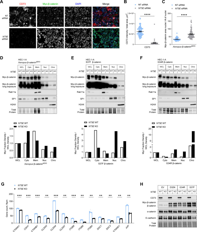
Figure 4. CD73 sequesters exon 3 mutant β-catenin to the cell membrane.
(A-C) HEC-1-A cells were transfected with Xenopus β-catenin∆EX3 and NT (non-targeting) or NT5E siRNA and cultured in 1% O2 and 5% CO2 for 48 hours. (A) Representative immunofluorescence images used in quantifying nuclear localization of myc-β-catenin∆EX3. Scale bar: 50 µm. Fluorescence intensity was determined with BZ-X800 Analyzer Macro cell count software (Keyence). (B) Validation of CD73 knockdown and (C) nuclear fluorescence intensity of myc-β-catenin∆EX3. (D-F) Representative immunoblots of n = 2 independent experiments of cellular fractionations from NT5E WT and NT5E KO HEC-1-A cells. Cells were transfected with (D) Xenopus β-catenin∆EX3 or patient-specific β-catenin mutants (E) S37F or (F) G34R. Graphs show densitometry for myc-β-catenin mutant expression for each cellular fraction normalized to myc-β-catenin mutant expression in the whole cell lysate (WCL). Cellular fraction markers: Rab11a (membrane), SP1 (nuclear), and H2AX (chromatin). (G) mRNA and (H) protein expression of differentially expressed cell-cell adhesion components in NT5E WT and NT5E KO HEC-1-A cells. Data represent the mean ± SEM. ****P < 0.0001, Mann-Whitney test; **P < 0.01, ***P < 0.005; Welch t-test.

