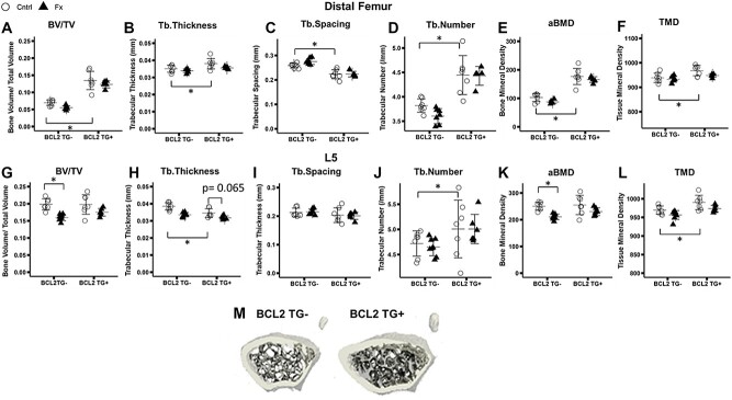
Figure 1.
Micro-computed tomography analysis of fifth lumbar vertebrae (L5) and contralateral (uninjured) distal femur trabecular bone. Group sizes: L5: TG- Cntrl: 7, TG- Fx: 8, TG+ Cntrl: 7, TG+ Fx: 6; distal femur: TG- Cntrl: 7, TG- Fx: 7, TG+ Cntrl: 7, TG+ Fx: 5. (A-F) Distal femur microarchitectural properties. Significant differences are evident between TG+ and TG- animals. (G-L) Lumbar spine trabecular microarchitectural properties. Significant decreases are seen in TG- fracture relative to control mice, but the same effect of fracture is not evident in TG+ mice (G-L). (m) Representative images of BCL2 TG- and TG+ distal femur metaphysis. *p < 0.05. Abbreviations: BCL2, B-cell lymphoma 2; TG-, non-transgenic; TG+, transgenic.

