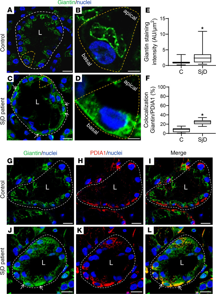
Figure 3. Increased Giantin protein levels in LSGs from patients with SjD.
(A–D) Representative images of Giantin (green) staining in LSG sections from individuals acting as controls and patients with SjD. (B and D) Higher-magnification images of epithelial cells surrounded by yellow dashed lines in A and C, respectively. (E) Giantin staining was quantified in acini from LSG sections of 5 individuals acting as controls and 5 patients with SjD. (F) Colocalization analysis of Giantin and PDIA1 in acini from LSG sections of 5 individuals acting as controls and 5 patients with SjD. Boxes represent the 25th–75th percentiles; the lines within the boxes represent the median; and the whiskers represent the minimum and maximum. *P < 0.05 was considered significant using the Mann-Whitney test. (G–L) Representative micrographs of double staining of Giantin (green) and PDIA1 (red) in a section of LSG from individuals acting as controls (G–I) and patients with SjD (J–L). Hoechst 33342 (blue) was used for nuclear staining. White arrows show Giantin staining in the basolateral region. The broken lines indicate acinar boundaries. L, lumen. Scale bars: 20 μm (A and C); 5 μm (B and D); 10 μm (G–I and J–L).

