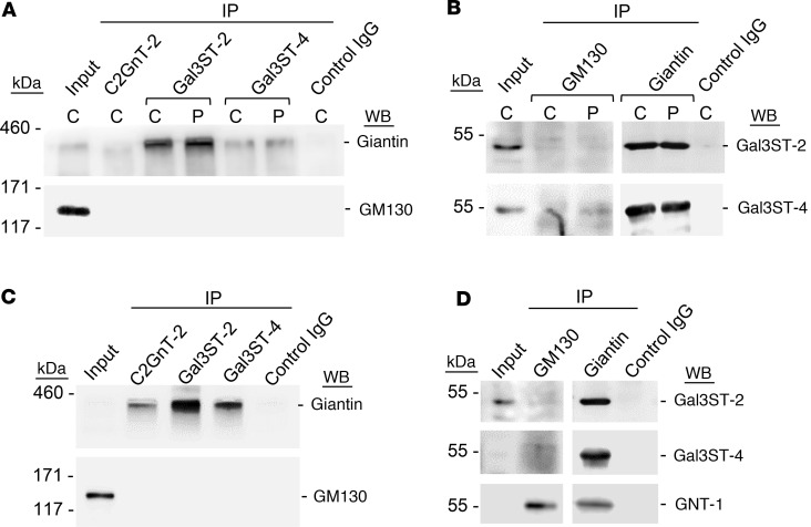
Figure 5. Formation of protein associations between Giantin and Gal3STs.
(A) C2GNT-2, Gal3ST-2, and Gal3ST-4 were immunoprecipitated (IP) from protein extracts of LSG from individuals acting as controls (C) and patients with SjD (P) and then analyzed by Western blot (WB) with anti-Giantin (~376 kDa) or anti-GM130 (130 kDa) antibodies. (B) GM130 and Giantin were IP from protein extracts of LSGs from individuals acting as controls and patients with SjD (P) and then analyzed by WB with anti–Gal3ST-2 (46 kDa) and anti–Gal3ST-4 (54 kDa) antibodies. (C) C2GNT-2, Gal3ST-2, and Gal3ST-4 were IP from protein extracts of HSG cells and then analyzed by WB with anti-Giantin or anti-GM130 antibodies. (D) GM130 and Giantin were IP from protein extracts of HSG cells and then analyzed by WB with Gal3ST-2 and Gal3ST-4 antibodies. The enzyme GNT-1 (57 kDa) was used as a positive control of IP GM130 and Giantin. For all experiments, the control IgG was α6 integrin.

