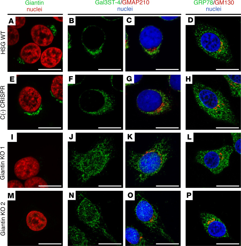
Figure 6. Altered Gal3ST-4 localization in Giantin-KO cells.
(A–D) Representative micrographs of WT HSG cells, (E–H) negative control [C(-)] CRISPR/Cas9 HSG cells, and (I–P) CRISPR/Cas9 Giantin-KO cells. A, E, I, and M show Giantin (green) and nuclei (red). B, F, J, and N show the green channel of the double staining of Gal3ST-4 (green) and GMAP210 (red). C, G, K, and O show the merged RGB channels. D, H, L, and P show the merged RGB channels of the double staining of GRP78 (green) and GM130 (red). Hoechst 33342 (blue) was used for nuclear staining. Scale bars: 10 μm.

