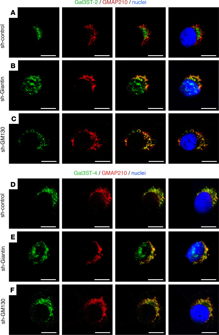
Figure 7. Altered Gal3ST-2 and Gal3ST-4 localization in sh-Giantin cells.
Representative micrographs of double staining of Gal3STs (green) and GMAP210 (red) in HSG cells. (A) In sh-control cells, Gal3ST-2 is located adjacent to the nucleus and partially colocalizes with GMAP210. (B) In sh-Giantin cells, Gal3ST-2 distribution changes, with diffuse staining “on the nucleus.” (C) Gal3ST-2 detection in sh-GM130 cells shows a distribution similar to sh-control cells. (D) In sh-control cells, Gal3ST-4 is located adjacent to the nucleus and partially colocalizes with GMAP210. (E) In sh-Giantin cells, Gal3ST-4 distribution changes, with diffuse staining “on the nucleus.” (F) Gal3ST-4 detection in sh-GM130 cells shows a distribution similar to that of sh-control cells. Hoechst 33342 (blue) was used for nuclear staining. Scale bars: 10 μm.

