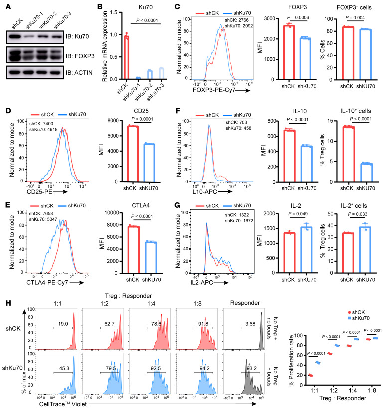
Figure 5. Human iTreg suppressive function is compromised upon knockdown of Ku70.
(A and B) Ku70 knockdown assay was performed in human iTregs using shRNA-Ctrl (shCK) or shRNA-Ku70 (shKu70) lentiviruses, and the expression levels of Ku70 and FOXP3 were examined by Western blot (A) and qRT-PCR (B). (C–E) Representative histogram of protein abundance of FOXP3, CD25, and CTLA4 in control and shKu70 knockdown human iTregs and summaries for the MFI. (F and G) IL-10 and IL-2 production from human iTregs were assessed after knockdown of Ku70 by lentiviruses carrying Ku70 shRNA. (H) In vitro suppression assay was performed in human iTregs with or without Ku70 knockdown. Human iTreg and CD8+ T cells (responder) were cocultured for 84 hours at a ratio of 1:1, 1:2, 1:4, or 1:8 with anti-CD3/CD28 beads for activation. The responder groups include 2 control groups: one without anti-CD3/CD28 bead stimulation and one with anti-CD3/CD28 bead stimulation but without coculture with Tregs. The generations of proliferating responder were monitored by dye dilution of CellTrace Violet (CTV). Data are representative of 3 independent experiments. Data are represented as means ± SD. Significance was measured by unpaired 2-tailed Student’s t test.

