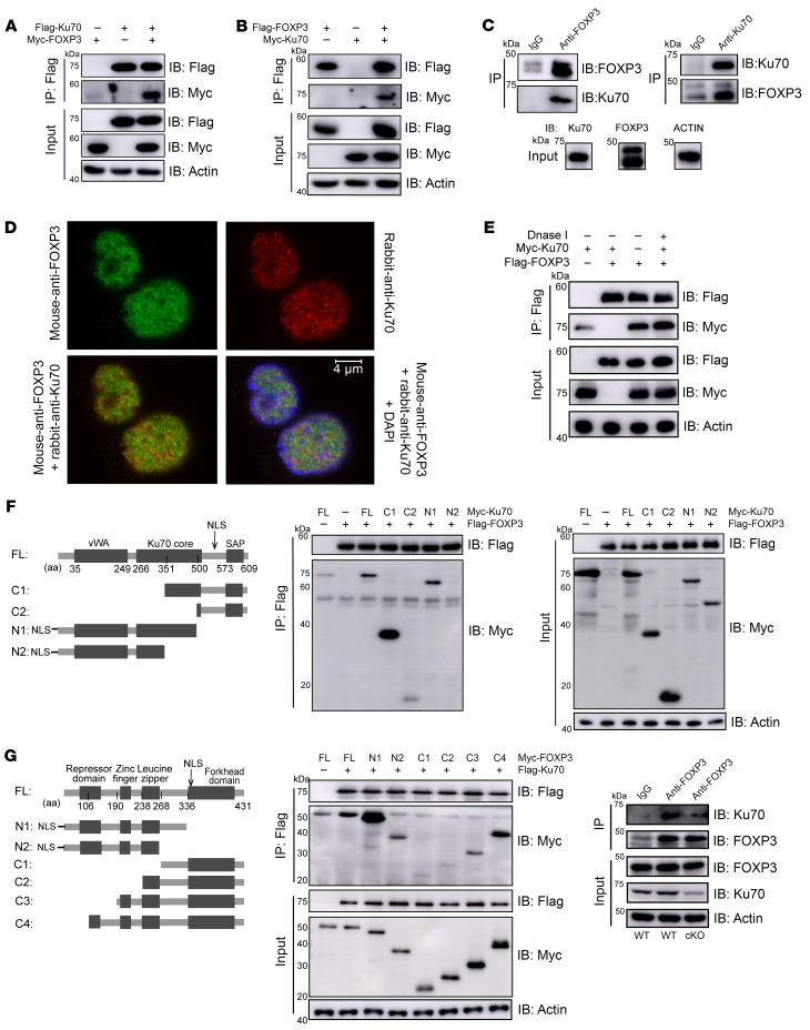
Figure 6. FOXP3 physically interacts with Ku70.
(A and B) HEK293T cells were transfected with Flag-Ku70/Myc-FOXP3 (A) or Flag-FOXP3/Myc-Ku70 (B). Cell lysate was IP with anti-Flag antibody plus protein A/G beads. The results were analyzed by Western blotting using the indicated antibodies. (C) Endogenous IPs with anti-Ku70 or anti-FOXP3 antibody were performed in human iTregs induced in vitro. The results were detected by Western blotting with indicated antibodies. (D) IF analysis of Ku70 (red), FOXP3 (green), and DAPI (blue) in human iTregs after 24-hour activation by anti-CD3/CD28 beads. (E) HEK293T cells were transfected with Flag-FOXP3/Myc-Ku70. DNase I (10 U) were added to HEK293T lysates, which were subjected to IP using anti-FLAG antibody followed by Western blotting. (F) Schematic representations of the plasmids encoding full-length (WT) and truncated mutants of Ku70 (left). Flag-tagged FOXP3 was cotransfected into HEK293T cells with the indicated Myc-tagged Ku70 constructs and IP with anti-Flag antibody followed by Western blotting. vWA, von Willebrand A domain; NLS, nuclear localization signal; SAP, SAF-A/B, acinus and PIAS domain. Ku70 mutant constructs include Ku70-C1 (amino acids [aa] 351–609), Ku70-C2 (aa 500–609), Ku70-N1 (aa 1–500), and Ku70-N2 (aa 1–351), with each fused to a Myc tag. (G) Schematic diagram of FOXP3 and the deletion constructs (left). Flag-tagged Ku70 was cotransfected into HEK293T cells with the indicated Myc-tagged FOXP3 constructs and IP with anti-Flag antibody followed by Western blotting. (H) Naive CD4+ T cells were sorted from spleen of WT or cKO mice (8 weeks old), then were induced into iTregs in vitro. Endogenous IPs with anti-Foxp3 or IgG antibody were performed after 5 days of induced differentiation. The results were analyzed by Western blotting using the indicated antibodies.

