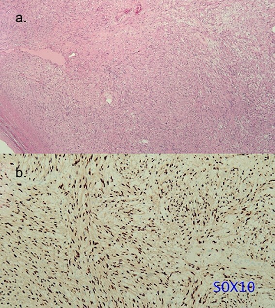
Figure 4.

Histopathology of the resected schwannoma. (a) Histologic analysis of the specimen revealing alternative cell-rich Antoni A type area and cell-poor Antoni B type area (hematoxylin and eosin stain). The resected specimen did not show pathological continuity between the left atrium and the tumor. (b) Tumor cells exhibiting intense positive staining for S-100 protein.
